Read the following summary of a primary research paper; then answer the questions below.
Skeletal myogenesis (the formation of muscle tissue) can he studied using in vitro cell cultures of the C2C12 myoblast cell line. C2C12 cells maintained in culture medium containing 20% fetal bovine serum (FBS) proliferate indefinitely, producing more myoblast. When these cells are deprived of FBS, however, they differentiate to form multinucleated myotubes that can no longer divide.
The differentiation of C2 C12 cells into myotubes is accompanied by changes in the expression of a number of proteins, including myosin heavy chain (MHC), p21 protein and several members of the MyoD family or muscle-specific transcription factors (e.g. MyoD and myogenin).
An experiment was carried out to investigate the order of some of the events that occur during differentiation of C2C 12 myoblast cell line in vitro. 1n this experiment, actively dividing C2C12 myoblasts were transferred from 20% FBS medium to differentiation medium (DM) containing only 2% FBS. Three to four days after the FBS concentration was reduced the majority of the myoblasts had differentiated into myotubes.
Antibodies that specifically recognise myogenin, p21 and MHC were used in immunofluorescence microscopy (Figure 1) and immunoblotting (Figure 2) to detect the expression of the proteins in the cultured C2C12 cells during differentiation.
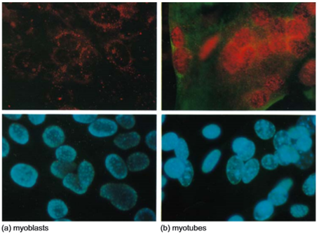
Figure 1 immunofluorescence antibody staining to reveal the expression and subcellular location of p21 and MHC in C2C12 myoblasts and myotubes. Cells were fixed then stained simultaneously wish three- stains: an anti-p21 antibody (detected with a secondary antibody carrying a green fluorescence signal), an anti-MHC antibody (detected with a secondary antibody carrying a green fluorescence signal) and a blue fluorescent stain that labels the cell nucleus. (a) (top and bottom images) shows undifferentiated C2C12 cells (myoblasts) growing in normal growth medium containing 20% FBS. (b) (top and bottom images) shows C2C12 multinuclear myotubes formed after 3 days growing in differentiation medium (DM) containing only 2% FBS. The cells have been photographed using different colour filters to detect different colours of fluorescence. In each case the top panel shows both the red and green fluorescence, while the bottom panel shows exactly the same cells with the blue nuclear slain, which reveals the position of every nucleus in the microscope field. Note that the very faint non-nuclear red staining in panel (a) top proved to be an irrelevant artefact caused by the non-specific sticking to cells of The secondary antibody carrying the red signal (a common problem with experiments involving antibodies).
In order to determine which of the C2C12 cells were still proliferating and which had differentiated, they were briefly incubated with bromodeoxyuridine (BrdU), an analog or the DNA base thymidine which only the actively dividing cells will incorporate into newly replicated DNA during the cell cycle (Figure 3), The incorporation of BrdU into DNA by actively dividing cells can be detected in the cell nucleus using a specific anti-BrdU antibody.
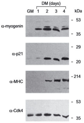
Figure 2 Immunoblot showing a lime course of expression of myo9enin, p21 and WHIC during C2C12 differentiation. Cell lysales were prepared from C2 12 cells maintained either in normal growth medium (GM) containing 20% FBS, or after 1 to 4 days growing in differentiation medium (DM) containing only 2% FES. Proteins in each cell lysate were separated by gel electrophoresis, and transferred to a membrane, which was then incubated with antibodies specific to myogenin, p21, MHC or Cdk4. The presence of each protein in the lysate can be observed as a discrete band, the intensity of which indicates the amount of the protein that is present. The protein Cdk4 is known to be expressed at the same level in both undifferentiated and differentiated C2C12 cells and provides a control proving that the same amount of cell extract was loaded in each lane. The position of migration of marker proteins of known molecular weight is shown to the right of the figure (kilodaltons, kDa).
Figure 3 BrdU incorporation into DNA to determine the percentage of the C2C12 cells that are psi actively dividing alter growth in differentiation medium for 1 day. (a) C2C12 cells were grown in differentiation medium (DM) containing 2% FBS for 1 day, then BrdU was aIDed to the cells for a 4-hour period, The cells were then fixed and stained simultaneously with anti-BrdU antibody (green, top panels) and either anti-myogenin antibody or anti-p21 antibody (red, bottom panels). The two left panels show two photos of the same cells; the lop one shows BrdU staining (green) and the bottom shows myogenin staining (red). The empty arrows indicate cell nuclei that are myogenin positive (Myog+) but BrdU negative (BrdU-), while solid white arrows indicate cell nuclei that arc both myogenin positive (Myog+) and BrdU positive (BrdU+). The two right-hand panels show two photos of another sample of cells; the top one shows BrdU staining (green) and the bottom shows p21 staining (red). The empty arrowheads indicate cell nuclei that are p21 positive (+) but BrdU negative (-). There are no nuclei that are both p21 positive (+) and BrdU positive (+) visible in this image. (b) A histogram showing the percentage of p21-positive cells (p21+) myogenin positive cells (Myog+), and cells that expressed neither p21 nor myogenin (p21-/Myog-) which were BrdU positive (BrdU+).
(a) When undifferentiated C2C12 cells are dividing in culture medium containing 20% FBS., which one of the following types of cell division are they undergoing:
(i) binary fission
(ii) mitosis
(iii) meiosis?
(b) Briefly explain why transferring cultured C2C12 cells into differentiation medium (DM) containing a reduced amount (2%) of fetal bovine serum (FBS) might trigger differentiation. (Your answer should be no more than 150 words.)
(c) In what order is the expression of the three proteins: MHC, myogenin and p21 switched on when C2C12 cells are transferred into differentiation medium (DM) containing 2% FBS? Explain clearly which of the data shown above brought you to your conclusion, and why. (Your answer should be no more than 200 words.)
(d) In which cell compartment are the myogenin and p21 proteins localised? Explain clearly which of the data shown above brought you to your conclusion and why, (Your answer should be no more than 200 words.)
(e) State the specific stage of the cell cycle in which undifferentiated C2C12 cells would incorporate BrdU.
(f) Approximately what percentage of each of the following classes of C2C12 cells is BrdU positive:
(i) p21-positive cells
(ii) myogenin-positive cells
(iii) cells which express neither p21 nor myogenin?
Does this data suggest that exit from the cell cycle (the point at which the differentiating C2C12 cells stop actively dividing) coins idea with the switching on of expression of myogenin, or of p21? Explain clearly how the data shown brought you to your conclusion, (Your answer should be no more than 200 words.)
(g) Briefly explain how the protein you identified in part (f) (either myogenin or p21) prevents cell division in the differentiating myoblasts. (Your answer should be no more than 200 words.)


 Jermaine Byrant
 Nicole Johnson