Neurology Take-Home
Question 1:
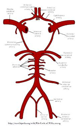
The Circle of Willis (cerebral arterial circle) is a vascular structure that allows confluence between the blood arriving from the left and right sides of the body through several arteries. After you have explored the anatomy of this structure, answer the following questions.
How does the organization of this structure help to perform its function(s)?
– The organization of this structure helps
In light of this function, what is the main potential advantage for the brain for such an organization?
– The Circle of Willis is an arterial anastomosis located in the brain. It unites both the brain’s anterior and posterior blood supply via the internal carotid and vertebral arteries. The loop that the Circle of Willis forms connects the anterior, posterior, left, and right blood supply of the brain. This is a huge advantage for the brain because it allows the blood supply to continue on another route if there is a blockage in an artery. Loss of bloody supply to tissue could be very damaging or life threatening. Without the Circle of Willis, a blocked artery could stop blood supply to a part of the brain resulting in a stroke or severe tissue damage.
According to A&P Revealed, the Circle is complete in only about 20% of individuals. Why might even an incomplete circle be better than no connection between sides at all in performing its main function?
– An incomplete Circle might be better than no connection between sides at all because even an incomplete Circle allows for some sort of collateral flow. Although not as effective as a complete Circle of Willis, no connection between sides at all would give alternative routes for blood to reach areas of the brain if an artery was blocked. An incomplete Circle still gives a chance that the blood will be able to take an alternate route to the brain in hopes of avoiding a complete blockage possibly resulting in a stroke.
Finally, undertakers find that the Circle of Willis complicates their task of embalming the body of a deceased person. How might this structure affect the ability to infuse the brain with embalming fluid, replacing all the blood in the vessels of the brain?
Hint: Think of a revolving door or a traffic circle.
– The purposeful structure, Circle of Willis, in a living person could complicate the task of embalming the body of a deceased person. The structure may make replacing all the blood in the vessels of the brain with embalming fluid difficult because the embalming fluid could be going around in the Circle instead of branching out into the anterior, posterior, left or right side of the brain. If the embalming fluid chooses the same route which avoids a particular area of the brain and continues in the circle similar to a round-a-bout, part of the brain will continue to have blood in the vessels instead of embalming fluid.
Question 2:
For the following description, identify FOUR (4) structures and pathways in the nervous system that are responsible for the experiences reported.
Specify whether each pathway is somesthetic or autonomic; and whether the response is reflexive or voluntary, cognitive or subconscious.
As they proceeded down the slope into the ghat, the rocks were slick and greasy.
Once, he lost his footing as the moist soil gave way.
– The pathway in the experience above is autonomic. Free nerve endings senses pain, heat, and cold. The afferent nerves that carry information from the sensory receptors to the brain are responsible. The response is reflexive and subconscious.
His arms swung out wildly as he arched his back and pumped his legs to regain his balance-the left leg flexing to lower the center of gravity and the right leg extending straight out into space.
– The response is reflexive and subconscious identified with autonomic pathway. The cerebral cortex is responsible for this experience, in particular the hypothalamus, and portions of the cerebellum and temporal lobe.
When he was fully upright, he sat against a rock to “catch his breath”.
When his work was done, he was grateful to be climbing back into the sunlight. Only a few feet to go.
His right leg was flexed at the knee and hip, and his left fully extended, Just as he transferred the weight from his left to his right foot, it happened.
– The pathway is somesthetic and the response is voluntary and cognitive. The somatic nervous system is involved as well as the motor cortex which triggers movement. The impulses are sent from the brain’s cerebral cortex to the muscles leading to the movement as indicated.
The soil gave way again under his right foot.
He jackknifed his body hard at the waist to change his center of gravity and reached out wildly with his right hand, grabbing onto a small bush just above his shoulder.
– This involves the motor impulses that control movements. In this case, the prefrontal cortex is involved which is responsible for control of emotions, planning, decision-making and self awareness. The pathways are therefore somesthetic and the response is voluntary and cognitive.
He was already tumbling down the side of the gully before he realized the pain caused by the four 2-inch long thorns that skewered his right palm. He hadn’t even known he was injured or consciously decided to let go of the bush. He only realized these things when he landed at the bottom of the gully with a loud thump.
Question 3:
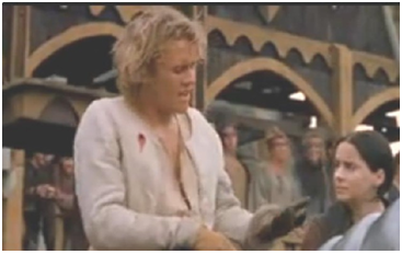
Now let us reconsider the case of Will Thatcher.
Using the material in muscle and nerve parts of the APR program – and combining it with other information from the course materials… describe the structure that is injured and how that damage may have caused the effects on his performance that you observed-especially his loss of grip and lower-arm control.
Hint: If you use the Layer 1 controls to fade down through the skin, you can use the surface features of the skin to compare the wound in the movie with the underlying anatomy.
– The structure that is injured is located in the upper right chest of Will Thatcher. The nerves located here extend from the brachial plexus and continue down the arm and into the hand. These major five nerves are the axillary, musculocutaneous, median, radial, and ulnar nerves. Each nerve serves a particular function in sending impulsesto and from a particular region of the arm and hand. Based on the nerves located in the shoulder region of the body, Will Thatcherwould not be able to flex or control his lower arm.
Which nerves are responsible the actions that Will found difficult to perform (Hint: Use the information in the Muscles section to identify the nerve supplies for various muscles whose functions Will lost).
Give your evidence for this conclusion.
– Will dropped the lance due to nerve damage in the shoulder that made him unable to grasp and keep hold of it. The muscles involved in the flexion of the digits and control of the lower forearm are flexor carpi ulnaris, flexor digitorum profundus, flexor digitorum superficialis, flexor pollicus longus, and pronator quadratus. The nerves that send motor signals to these muscles are the ulnar nerve and median nerve, which are located in the upper quadrant of the shoulder. Will did not acquire complete breakage of these individual nerves, so we infer that the injury was more proximal towards the root of where these nerves stem, which is the brachial plexus. The medial cord of the brachial plexus branches into the ulnar nerve, contributes to the median nerve, and helps with the control of the forearm.
Which nerves are associated the actions that Will still could perform?
Give your evidence for this conclusion.
– In the video clip, we see that Will was still able to somewhat flex his radioulnar joint. The muscles that are responsible for this action are the brachialis, biceps brachii, and brachioradialis, which are controlled by the musculocutaneous and radial nerves, which were not damaged as much in the injury as the other nerves. The lateral and posterior cords of the brachial plexus must not have been severed because these two cords brach out to the musculocutaneous and radial nerves.
Summarize your conclusions about the nature of the injury, and explain how the evidence in your answers above led you to your conclusions about the nature of this injury and why it produced the problems that it did for our hero.
– Will acquired an injury in his upper right chest, which severely damaged the medial cord of the brachial plexus, which greatly affected his ulnar and median nerve function. We see in the clip that he cannot flex his hand or fingers in order to grasp the lance, and he cannot control parts of his lower arm, which leads us to conclude that the root of the two prior nerves was damaged. He was able to flex his elbow, which leads us to conclude that the posterior and lateral cords of the brachial plexus that branch into the musculocutaneous and radial nerves was not severely injured compared to the medial cord of the brachial plexus.
Question 4:
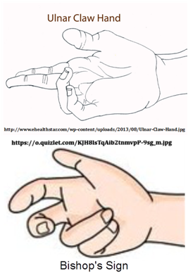
Two different types of peripheral nerve damage produce similar hand deformities. One is called the “Bishop’s sign” or “benediction” because of the similarity to the way that clerics position their hands when blessing a congregation. The other is sometimes called the “ulnar claw”. These occur when nerve damage affects the relative positions of the fingers in similar ways, but for different reasons.
Examine the neuromuscular interactions that are responsible for these two phenomena. How do the nerves, muscles, bones, and joints interact in making these hand signs different from each other and from the unmodified positions of these structures in the hand?
– The ulnar claw is formed from damage to the ulnar nerve at the wrist and affects the little and ring fingers of the hand. These fingers are hyperextended at the metacarpophalangeal joint and flexed at both interphalangeal joints. The muscles paralyzed are the medial two lumbricals.
– The bishop’s sign is formed from damage to the median nerve at the elbow and affects the miIDle and index fingers. The bishop’s sign is only apparent while making a fist and the muscles paralyzed are the lateral half of flexor digitorum profundus and the lateral two lumbricals.
Be sure to identify the muscles involved in these conditions and the nerves that supply them, and to describe how the interactions of the nerves with the muscles, bones, and joints accomplish this phenomenon.
– Anatomically speaking, explain how these two conditions (what nerves supply the “normal/natural” actions of these muscles and how their activation must be changed to produce these movements of the bones and joints).
– In aIDition, identify where the “motor program” is primarily affected … at the level of the hand muscles, in the PNS, in the spinal cord, or in the brain (and where in the brain)..so that these signs are produced.
HINT: remember that this assignment is in the unit on the brain and nervous system, so your answers should be focused accordingly.


 Jermaine Byrant
 Nicole Johnson